
- What anatomy and physiology are, and eight ways they help in exercise and sports
- The parts and functions of the skeletal system, and how bones are classified by tissue and by shape
- How joints are classified by function and by structure, and the six types of synovial joints with sports examples
- The three types of muscle, the four properties of skeletal muscle and the six functions of muscles
- The structure of the heart and the pulmonary, systemic and coronary circulations
- The parts of the respiratory system in order, and how the diaphragm helps us breathe
In the CBSE Physical Education (048) curriculum for Class XI, this is Unit 7: Fundamentals of Anatomy and Physiology in Sports, worth 8 marks in the 70-mark theory paper. The prescribed book is CBSE’s own Physical Education Class XI textbook (2nd edition, 2023); there is no NCERT book for this subject. The curriculum lists five topics: the definition and importance of anatomy and physiology; functions of the skeletal system, classification of bones and types of joints; properties and functions of muscles; structure and functions of the circulatory system and heart; and structure and functions of the respiratory system. All five are covered below.
1 Anatomy and Physiology
A sports coach needs to know two things about the human body: what it is made of and how it works. The first is anatomy, the second is physiology. Together they tell us why a tall player suits basketball, why a helmet is shaped the way it is, and why the heart beats faster when we run.
1.1 Anatomy
Anatomy is the science that deals with the structure of the body and the relationship between the body parts. In other words, it is the study of the shape and structure of the human body and its parts, along with their relationship to one another.
- The word comes from two Greek words: ana = apart and tomy = to cut. Early physicians and scientists dissected (cut open) dead bodies to see how the parts were joined. So anatomy can also be defined as the science of the structure of a body learned by dissection.
Gross (macroscopic) anatomy
- Studies the large structures of the body
- Can be seen with the naked eye
- Example: the digestive system
Microscopic anatomy
- Studies structures that are too small to see
- Needs a microscope
- Example: the cells of the human body
- The study of tissues under the microscope is called histology
1.2 Physiology
Physiology is the scientific study of the functions of human beings. Or, physiology is the study of the responses of the human body to physical activity.
- The word comes from the Greek physio = nature and logio = the study of.
- It covers the detailed working of the body and its parts, and how the body responds to a stimulus. When we walk, all body parts work together in a synchronised way, and the heart keeps working to supply oxygen and other nutrients to our cells. Studying this is physiology.
- Physiology is the detailed study of life, from the working of the smallest cells to tissues and whole organisms.
| Branch of physiology | What it studies |
|---|---|
| Human physiology | One specific organism: the human being |
| Cellular physiology | The function of cells |
| Systemic physiology | The function of the body’s systems (such as the circulatory or respiratory system) |
| Point | Anatomy | Physiology |
|---|---|---|
| Question it answers | “What is it and where is it?” | “How does it work?” |
| Studies | Structure, shape and relationship of body parts | Functions of body parts and the body’s responses to activity |
| Greek root | ana (apart) + tomy (to cut) | physio (nature) + logio (study of) |
| Example | The heart has four chambers | The heart beats faster when we run |
2 Importance of Anatomy and Physiology in Exercise and Sports
The book gives eight ways in which knowing the body helps a person in physical activity and sports.
2.1 The eight points
Helps in physical fitness
Knowing how the body works helps a person get a fit and healthy body, for example by building muscle strength and muscle endurance through the right exercises.
Knowledge of body structure
We can assess our strengths and weaknesses and improve the body, for example by designing an exercise routine that suits a person’s needs and body structure.
Knowledge of the functions of organs
Knowing how the body’s systems work helps us train them to work at their best. Knowing the cardiovascular system shows the value of the heart and why activity keeps it strong.
Helps in selection of games
A game can be chosen to suit the body structure. A tall person suits basketball or volleyball; kho-kho suits a person of short height.
Protects from sports injuries
Sprain, contusion, fracture and dislocation are common. Protective equipment is designed from knowledge of bones, muscles, tendons and ligaments, for example cricket leg pads and helmets.
Helps in rehabilitation
It helps an injured player recover from soft or hard tissue injury and reach the pre-injury level. If a friend twists an ankle, you can give first aid such as ice before going to the doctor.
Helps maintain a healthy body
Lifestyle changes and knowledge of the body help us reach an ideal weight. Anatomy tells us about good and bad posture while sitting, standing, lying down and running.
Individual differences between male and female athletes
Male and female bodies differ, so equipment differs too. The difference in the structure of the shoulders is why the shot put, discus, hammer and javelin have different weights for men and women.
“Fit Body Organs Select Safe Recovery, Healthy Individuals”: Fitness · Body structure · Organs · Selection · Safety from injury · Rehabilitation · Healthy body · Individual differences
3 The Skeletal System
The human skeletal system is the internal framework of the body. It consists of bones, cartilages, joints and ligaments (with the tendons that tie muscles to it).
3.1 What makes up the skeleton
| Part | What it is | What it does |
|---|---|---|
| Bones | The rigid part of the skeleton | Support the body; their shapes suit different jobs (metacarpals and phalanges let the hand form a fist) |
| Cartilage | Semi-rigid yet flexible connective tissue, more flexible than bone | Gives shape and flexibility: shapes the ears and nose, and lets the chest expand while breathing |
| Tendons | Strong bands of fibrous connective tissue | Connect a muscle to a bone |
| Ligaments | Fibrous cords | Connect a bone to another bone at a joint |
| Joints | Points where two or more bones meet | Permit movement |
Joint: a point where two or more bones are connected in the body in a manner that permits movement.
Cartilage: a form of connective tissue that is semi-rigid yet flexible, found in the joints and in places such as the nose, throat and ears.
Tendon: a strong piece of tissue connecting a muscle to a bone.
Ligament: fibrous cords that bind the bones together at joints.
Tendon = muscle To bone · Ligament = Links bone to bone
3.2 Axial and appendicular skeleton
Axial skeleton
- The central axis of the body
- Skull, vertebral column (the vertebrae), rib cage (ribs and sternum)
- Mainly protects organs: brain, spinal cord, heart and lungs
Appendicular skeleton
- Attached to the axial skeleton
- Shoulder girdle, pelvic girdle, and bones of the upper and lower limbs
- Mainly allows movement of the arms and legs

General facts that the book’s case study asks about (not printed in the unit’s text): the femur (thigh bone) is the longest bone; there are 8 carpals in each wrist; the vertebral column has 33 vertebrae in a child, which become 26 bones in an adult because the sacral and coccygeal vertebrae fuse into the sacrum and coccyx.
3.3 Functions of the skeleton
Shape and support
The skeleton gives the body its shape and holds it up.
Movement
It forms the framework to which muscles are attached. Movement happens when the muscles contract and relax and pull on the bones.
Protection
It protects the soft internal organs: the ribcage protects the heart and lungs; the skull protects the brain.
Storehouse of minerals
The hard substance of the bones stores minerals such as calcium.
Formation of blood cells
Blood cells are formed inside the cavities of the bones (in the bone marrow). This is called haemopoiesis.
Haemopoiesis (also written haematopoiesis) is the formation of blood cells within the cavities of the skeleton.
4 Classification of Bones
Bones are classified in two ways: by the tissue they are made of, and by their shape and size.
4.1 By bone tissue
Compact bone
- Dense, smooth and homogenous (the same all through)
- Forms the hard outer layer of bones
Spongy bone
- Made of needle-like pieces of bone with spaces between them
- Found inside bones, for example in the core of short bones and between the layers of flat bones
4.2 By shape and size
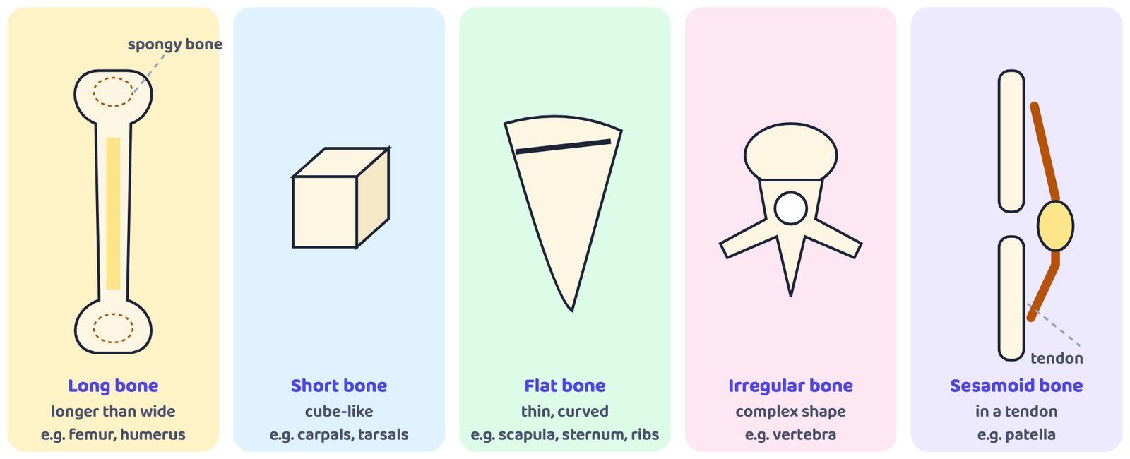
| Type | Shape and make-up | Job | Examples |
|---|---|---|---|
| Long bones | Cylindrical; more length than width (named for their shape, not their size). Covered with a fibre sheet, and with a thin sheet of cartilage where they join another bone. Contain yellow and red bone marrow. | Hard and dense; give strength, structure and mobility. Their red marrow produces blood cells. | Humerus, radius, ulna (arm); femur, tibia, fibula (thigh and leg); metacarpals; phalanges of fingers and toes |
| Short bones | Cube-like: length, width and thickness about equal. A core of spongy bone covered by a thin layer of compact bone. | Limited motion; they glide on one another | Carpals (wrist), tarsals (ankle); the book pictures a carpal (the capitate) and the talus |
| Flat bones | Thin and usually curved. A central layer of spongy bone between two layers of compact bone. | Form a bony cage that protects soft internal organs | Cranial bones, ribs, sternum, scapula, hip bone |
| Irregular bones | Vary in shape and structure; do not fit the other groups; complex shape | Protect internal organs; the vertebrae protect the spinal cord | Vertebrae; some bones of the skull |
| Sesamoid bones | Small, round bones embedded in tendons, shaped like a sesame seed. Their number varies from person to person. | Sit inside a tendon at a joint | Patella (kneecap), the example the book gives |
In a “name the type of bone” question, think of the shape first: long (femur), cube (carpal), plate (scapula), odd shape (vertebra), seed in a tendon (patella). The femur is a long bone, the patella is a sesamoid bone, the radius is a long bone and a metatarsal is a long bone too.
5 Joints
A joint or articulation is the point where two or more bones meet and muscles act on them to cause movement.
Most joints move, but not all of them. Some joints allow only limited movement and some are completely immovable. Joints are classified in two ways: by function (how much they move) and by structure (what holds the bones together).
5.1 Functional classification
The functional classification is based on the amount of movement the joint permits.
| Type | Also called | Where mostly found |
|---|---|---|
| Synarthroses | Immovable joints | Axial skeleton, where protection of internal organs and firm attachment matter most |
| Amphiarthroses | Slightly movable joints | |
| Diarthroses | Freely movable joints | Limbs, where movement and mobility matter most |
5.2 Structural classification
The structural classification depends on whether fibrous tissue, cartilage or a joint cavity separates the bones at the joint. Fibrous joints are generally immovable, synovial joints are freely movable, and cartilaginous joints include both immovable and slightly movable joints.
(a) Fibrous joints
The bones are united by fibrous tissue and show little or no movement. There are three kinds:
| Kind | How the bones are held | Example |
|---|---|---|
| Sutures | A tight union that prevents any movement. Found only between the bones of the skull (cranium). A foetus’s skull bones are unfused; after birth they slowly fuse and become immovable to protect the brain from impact. | Bones of the skull |
| Syndesmoses | Bones separated by some distance and united by ligaments. Because these joints are not flexible, ligament injuries are common here, particularly at the wrist and ankle. | Fibrous membrane joining the lower (distal) ends of the radius and ulna |
| Gomphoses | A peg fitted into a socket and held by ligaments | A tooth in its socket |
(b) Cartilaginous joints
Two bones are united by cartilage. Very slight movement can occur. The bone surfaces are connected by pads (discs) of fibrocartilage. Examples: the cartilage of growing long bones, and the cartilage between the ribs and the sternum.
(c) Synovial joints
These are freely movable joints, mostly found in the limbs. They contain synovial fluid, which lets the bone ends move smoothly. All synovial joints have four distinguishing features:
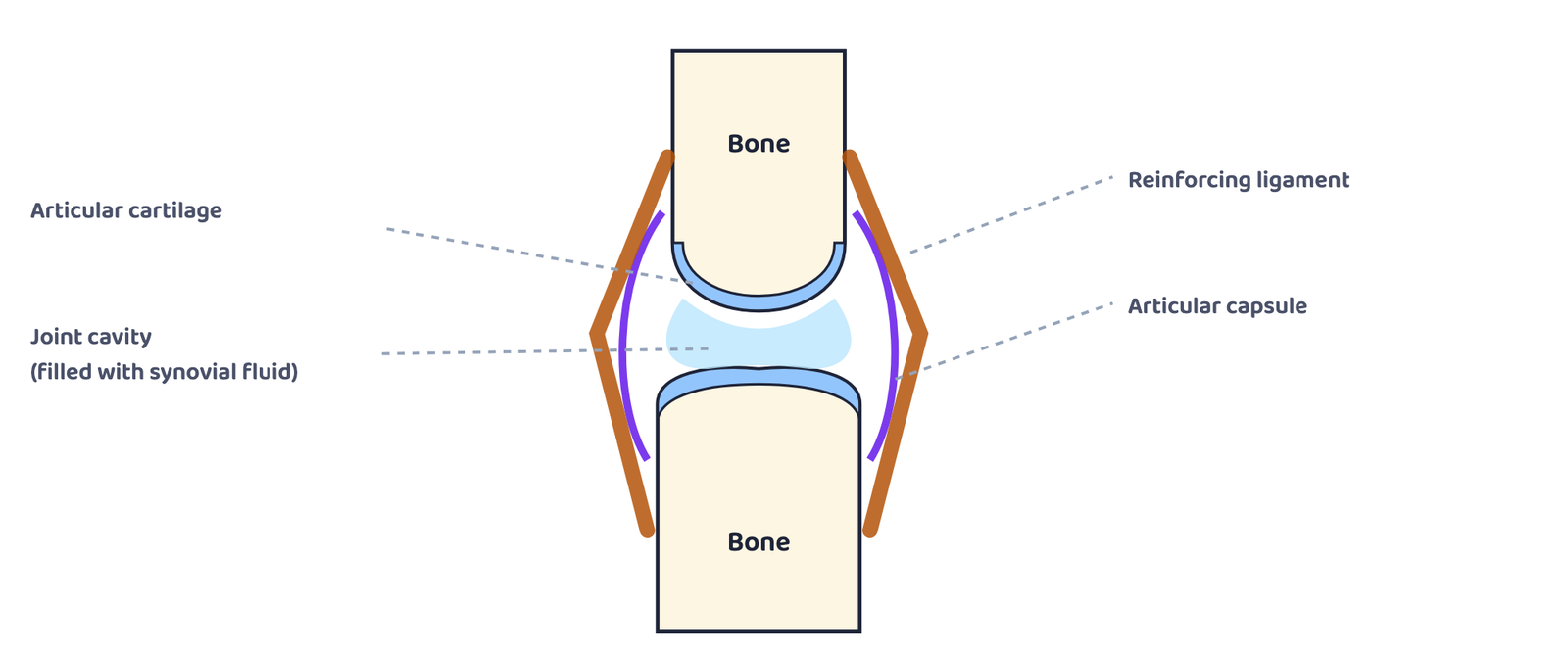
Articular cartilage
Smooth cartilage covering the ends of the bones that meet.
Articular capsule
The sleeve that encloses the joint.
Joint cavity
The space between the bones, filled with synovial fluid.
Reinforcing ligament
Ligaments that strengthen the joint and hold the bones together.
5.3 Types of synovial joints
Synovial joints are classified by the shape of the articulating (meeting) surfaces. There are six types.
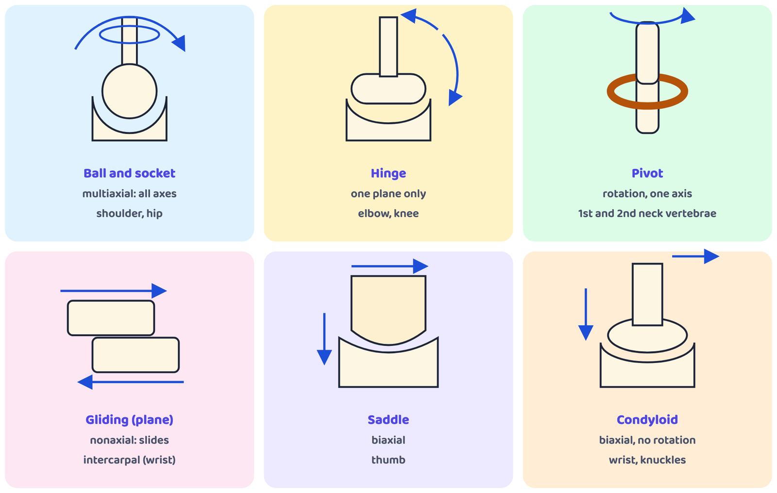
| Type | How the bones fit | Movement allowed | Examples | Use in sport |
|---|---|---|---|---|
| Ball and socket | Ball-shaped head of one bone fits into a cup-like socket of another | Greatest range; multiaxial: movement in all axes, including rotation | Hip joint, shoulder joint | Overhead clear in badminton; bowling in cricket |
| Hinge | Cylindrical end of one bone fits into a trough-shaped surface of another | Back and forth, like a door hinge; angular movement in one plane only (flexion and extension) | Elbow, knee | Flexion and extension build the biceps, triceps and quadriceps |
| Pivot (rotary) | The moving bone rotates within a ring formed by a second bone and a ligament | Only rotation around a single axis | Joint between the first and second cervical (neck) vertebrae | Turning the head from side to side |
| Gliding (plane) | Flat or nearly flat surfaces meet | Bones glide past each other in any direction (up and down, left and right, diagonally); nonaxial: no rotation | Intercarpal joints (joints of the wrist) | Gliding of the wrist bones on one another |
| Saddle | Surfaces shaped like a saddle, with both convex and concave areas | Rock back and forth and side to side; biaxial; limited rotation | Thumb joint | A thumb war |
| Condyloid | Egg-shaped surface of one bone fits into an oval cavity of another | Side to side and back and forth, but no rotation; biaxial | Wrist joint; metacarpophalangeal (knuckle) joints | Using the wrist to dribble the ball in basketball |
“Big Hungry Pigs Grab Six Carrots”: Ball and socket · Hinge · Pivot · Gliding · Saddle · Condyloid
Match the joint to the movement: rotating the head = pivot; bending the elbow (flexion) or straightening it (extension) = hinge; swinging the arm or leg in a circle (circumduction), or raising the arms sideways (abduction) and lowering them (adduction) = ball and socket at the shoulder or hip.
6 Muscles: Types, Properties and Functions
All movement in our body is done by the contraction (shortening) or relaxation of muscles. Movement can be:
- Voluntary: under our control, such as running or walking.
- Involuntary: not under our control, such as the pumping of blood by the heart.
6.1 Types of muscle tissue
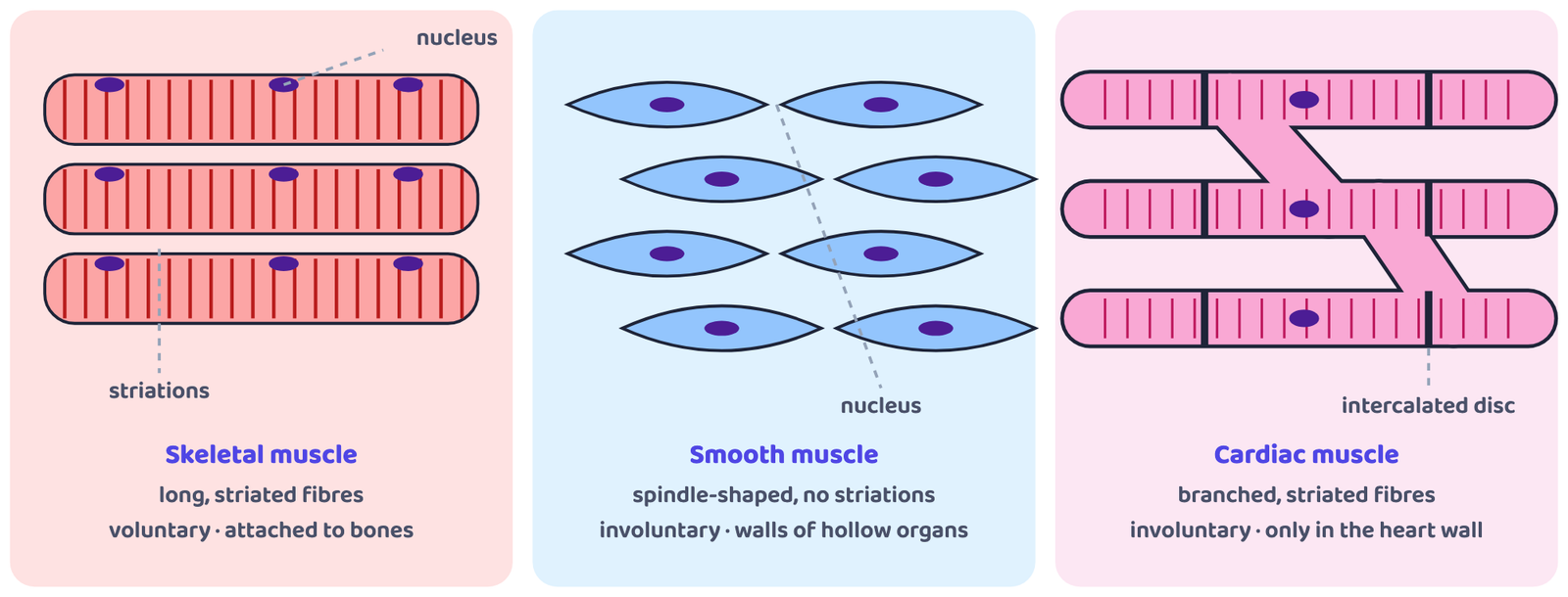
| Feature | Skeletal muscle | Smooth muscle | Cardiac muscle |
|---|---|---|---|
| Where found | Attached to the skeleton by tendons | Walls of hollow organs: stomach, urinary bladder, respiratory passages; also in the eyes and the skin | Only in the heart, where it forms the walls |
| Striations (stripes) | Yes: also called striated muscle | No striations | Yes, but not as clearly as skeletal muscle |
| Shape of cells | Long fibres | Elongated, slender, spindle-shaped | Long, striated (branched) fibres |
| Control | Voluntary: we control them at will | Involuntary | Involuntary |
| Speed of contraction | Fastest, but tire if they work for long | Slowest | In between the two |
| Examples of work | Walking, running, smiling, eating; exerting force | Changing the size of the iris and the shape of the lens; making hair stand on end in cold or fear | Pumping blood all life long |
6.2 Properties of skeletal muscle
Skeletal muscle has four major functional properties.
Excitability is the ability to respond to a stimulus, which may come from a motor neuron (nerve) or a hormone.
Contractility is the ability of muscle cells to shorten themselves forcefully (self-contraction).
Extensibility is the ability of a muscle to stretch, or its capacity to lengthen.
Elasticity is the ability of a muscle to recoil, or bounce back to its original length after being stretched.
“Every Champion Exercises Easily”: Excitability · Contractility · Extensibility · Elasticity
6.3 Functions of muscles
Movement
Muscles give the body strength; their contraction and relaxation produce every movement: gross movements like walking and fine movements like writing.
Maintenance of posture
Muscles hold our posture while sitting or walking. Strong muscles give good posture; weak muscles cannot hold it.
Heat generation
Muscles help keep body temperature. When it falls, skeletal muscles start contracting to bring it back to normal: this is shivering.
Respiration
The diaphragm, a muscle below the lungs, contracts so that the chest cavity gets bigger and air fills the lungs; when it relaxes, air is pushed out.
Constriction of organs and vessels
Smooth muscle contractions move food through the digestive tract, pass urine out of the body and push secretions out of glands.
Pumping blood
The heart pumps blood, and the smooth muscles in the walls of arteries and veins help move blood between the heart and the cells.
7 The Circulatory System and the Heart
The circulatory system is responsible for transporting blood: oxygenated blood from the heart to the body cells and deoxygenated blood back to the heart, and then deoxygenated blood from the heart to the lungs and oxygenated blood back to the heart, through arteries and veins.
7.1 Parts of the circulatory system
| Part | Details |
|---|---|
| Heart | Four chambers: two atria (receiving chambers, on top) and two ventricles (pumping chambers, below). A thick muscular wall, the septum, separates the right and left sides. |
| Blood vessels | Arteries take blood away from the heart to the cells; veins bring blood back to the heart; capillaries are the thinnest vessels, where nutrients and oxygen are exchanged. |
| Blood | The body fluid that carries nutrients and oxygen. It contains red blood cells, white blood cells, plasma and platelets. It is red because of the pigment haemoglobin. |
7.2 The heart
The heart is made of cardiac muscle. It contracts and relaxes in turn all through life.

| Chamber | Receives blood from | Sends blood to |
|---|---|---|
| Right atrium | The body, through the veins (vena cava): deoxygenated | Right ventricle |
| Right ventricle | Right atrium | The lungs, through the pulmonary artery |
| Left atrium | The lungs, through the pulmonary veins: oxygenated | Left ventricle |
| Left ventricle | Left atrium | The whole body, through the aorta |
Systole is the contraction of the heart, when the ventricles pump blood out of the heart.
Diastole is the relaxation of the heart, when its chambers fill with blood.
Stroke volume is the amount of blood pumped out of a ventricle in each contraction.
Cardiac output is the amount of blood pumped out of the heart in one minute.
Stroke volume is per beat; cardiac output is per minute. So cardiac output = stroke volume × number of beats in a minute (heart rate). When you run fast, the heart beats faster to meet the body’s need for oxygen, so cardiac output rises.
7.3 Functions of the heart
- Pumps oxygenated blood to every part of the body through the aorta (systemic circulation).
- Pumps deoxygenated blood to the lungs through the pulmonary artery to collect oxygen (pulmonary circulation).
- Receives blood back from the body (right atrium) and from the lungs (left atrium).
- Keeps the two kinds of blood apart with the septum.
- Changes its rate to suit the body’s needs: it beats faster during exercise.
- Supplies its own muscle with blood through the coronary circulation.
7.4 Three parts of the circulation
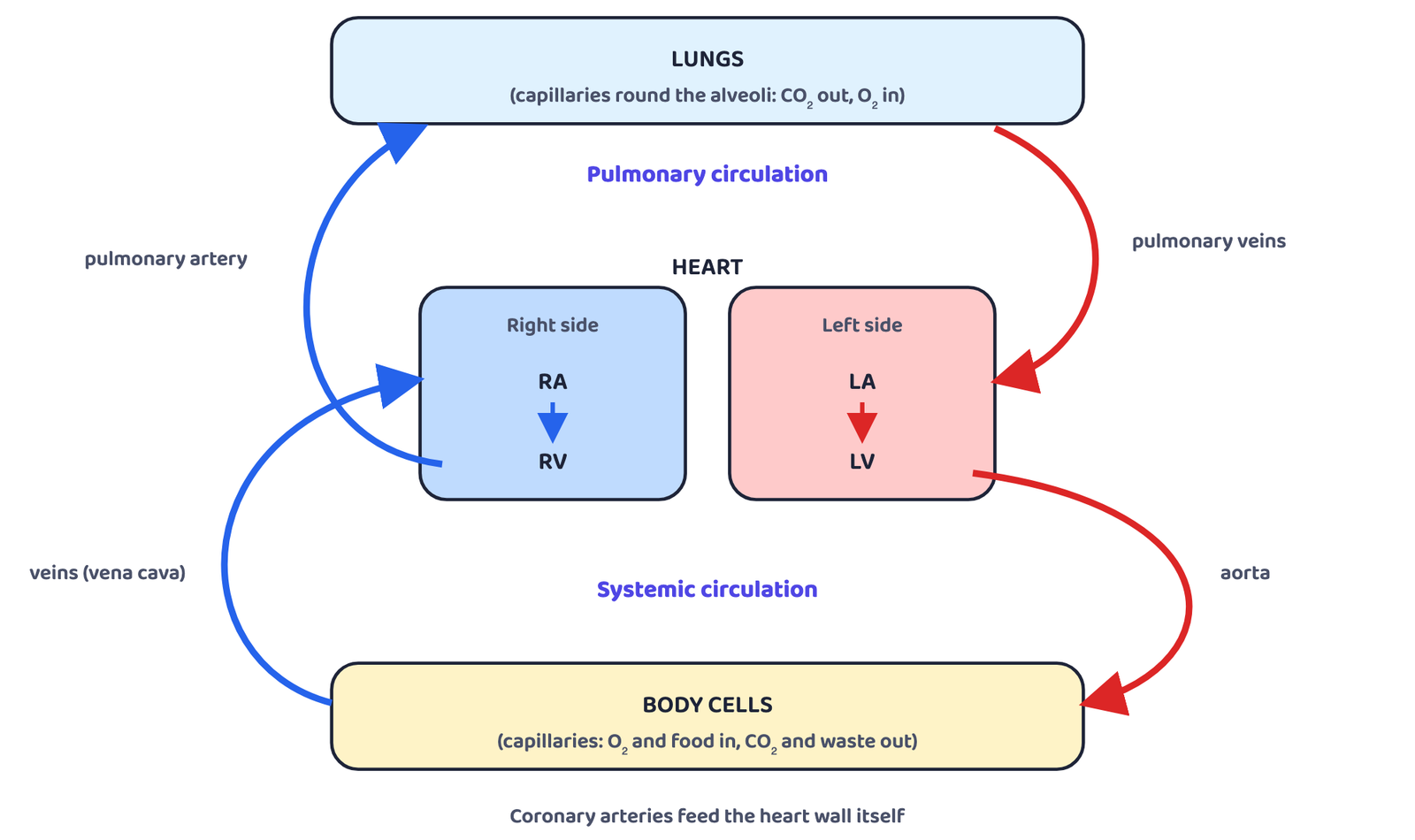
(a) Pulmonary circulation (heart → lungs → heart)
The pulmonary artery is the only artery that carries deoxygenated blood, and the pulmonary vein is the only vein that carries oxygenated blood. Both belong to the pulmonary circulation.
(b) Systemic circulation (heart → body → heart)
Both circulations happen at the same time, and the heart controls the flow to suit the body’s needs.
(c) Coronary circulation
The heart works without rest from birth to death, so its own muscle also needs oxygen. Coronary arteries carry oxygenated blood to the heart muscle, and coronary veins take the deoxygenated blood back to the right atrium.
7.5 Blood vessels
Arteries
- Carry blood away from the heart
- Carry oxygenated blood (except the pulmonary artery)
- Example: aorta
Veins
- Carry blood back to the heart
- Carry deoxygenated blood (except the pulmonary veins)
- Example: vena cava
8 The Respiratory System
Respiration is made up of two phases: inspiration, when we inhale (breathe in) oxygen, and expiration, when we exhale (breathe out) carbon dioxide.
8.1 Processes of respiration
8.2 Structure of the respiratory system
Air passes through these parts in order:
The lungs hold the bronchi, bronchioles and alveoli, and the diaphragm below them powers breathing. The book lists ten parts: nose, nasal cavity, pharynx, larynx, trachea, bronchi, lungs, bronchioles, alveoli and diaphragm.

| Part | Structure and function |
|---|---|
| Nose | The visible structure on the face, together with the internal nasal cavity behind it. |
| Nasal cavity | Extends from the opening of the nose to the pharynx; divided into right and left sides by the nasal septum. |
| Pharynx | The common passageway of both the digestive and respiratory systems (food and air). Has three regions (table below). |
| Larynx (voice box) | An outer casing of nine cartilages connected by muscles and ligaments. |
| Trachea (windpipe) | A membranous tube of connective tissue and smooth muscle. |
| Bronchi | The trachea divides into the left and right primary bronchi. They give air a passage into and out of each lung; their mucous membrane traps debris and pathogens (germs). |
| Bronchioles | About 1 mm across; branch further into tiny terminal bronchioles (more than 1000 in each lung) that lead to the gas-exchange structures. Their muscular walls have no cartilage, so they can change the size of the tube to increase or decrease airflow. |
| Alveoli | An alveolar duct (a tube of smooth muscle and connective tissue) opens into a cluster of alveoli. An alveolus is one of the many small, grape-like sacs; gases are exchanged here with the blood in the surrounding capillaries. |
| Lungs | The principal organs of respiration: spongy, pinkish, like two upside-down cones in the chest. The right lung has three lobes; the left lung has two lobes, to make room for the heart. Each lung is covered by a membrane called the pleura. |
| Diaphragm | A thin skeletal muscle that separates the abdomen from the chest. It powers breathing (Section 8.3). |
| Region of the pharynx | Extends from | Carries |
|---|---|---|
| Nasopharynx (upper part) | Internal nares (openings) of the nasal cavity to the level of the uvula | Air |
| Oropharynx | Uvula to the epiglottis; bordered above by the nasopharynx and in front by the mouth (oral cavity) | Air and food |
| Laryngopharynx | Epiglottis to the lower margin of the larynx, where the digestive and respiratory paths separate | Air and food |
8.3 How the diaphragm helps us breathe
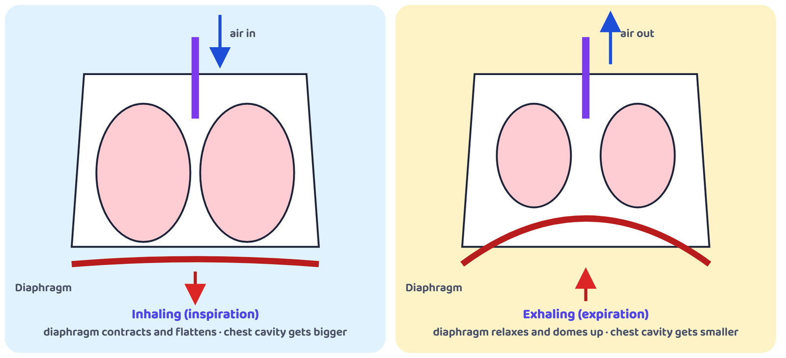
Inhaling (inspiration)
- The diaphragm contracts and flattens
- The chest cavity gets bigger
- This creates a vacuum effect that pulls air into the lungs
Exhaling (expiration)
- The diaphragm relaxes and moves up
- The chest cavity gets smaller
- Air is pushed out of the lungs
8.4 Functions of the respiratory system
- Ventilation: moves air into and out of the lungs.
- Gas exchange: supplies oxygen to the blood and removes carbon dioxide from it, in the alveoli (external respiration); the blood then exchanges gases with the tissues (internal respiration).
- Transport of oxygen and carbon dioxide in the blood, working with the circulatory system.
- Protection: the mucous membrane of the bronchi traps debris and pathogens.
- Control of airflow: the muscular walls of the bronchioles change the size of the tubes.
- Voice: the larynx is the voice box.
Gas exchange happens in the alveoli, not in the diaphragm. The diaphragm is the muscle that makes the chest bigger and smaller so that air moves in and out.
- Anatomy = structure (ana + tomy); physiology = function (physio + logio). Gross vs microscopic anatomy; human, cellular and systemic physiology.
- Importance (8): fitness, body structure, organ functions, selection of games, injury protection, rehabilitation, healthy body, male-female differences.
- About 300 bones at birth, 206 in adults. Axial: skull, vertebral column, rib cage. Appendicular: girdles and limbs.
- Functions of the skeleton: shape and support, movement, protection, mineral store, haemopoiesis.
- Bones by tissue: compact, spongy. By shape: long (femur), short (carpals), flat (scapula), irregular (vertebra), sesamoid (patella). Shortest: stapes.
- Joints by function: synarthroses, amphiarthroses, diarthroses. By structure: fibrous (sutures, syndesmoses, gomphoses), cartilaginous, synovial.
- Synovial joint features: articular cartilage, articular capsule, joint cavity, reinforcing ligament. Types: ball and socket (shoulder, hip), hinge (elbow, knee), pivot (neck), gliding (intercarpal), saddle (thumb), condyloid (wrist).
- Muscles: skeletal (striated, voluntary, 40% of body weight), smooth (spindle, involuntary, slowest), cardiac (heart only, involuntary). Properties: excitability, contractility, extensibility, elasticity.
- Heart: 2 atria + 2 ventricles, septum. Right ventricle → pulmonary artery → lungs → pulmonary veins → left atrium → left ventricle → aorta → body → veins → right atrium.
- Respiratory path: nose → nasal cavity → pharynx → larynx → trachea → bronchi → bronchioles → alveoli. Right lung 3 lobes, left lung 2. Diaphragm contracts and flattens to breathe in.
- Can I define anatomy and physiology and write the eight points of their importance in sports?
- Can I list the five functions of the skeleton and the five types of bones with examples?
- Can I classify joints both ways and describe the six synovial joints with examples?
- Can I compare skeletal, smooth and cardiac muscles and explain the four properties of skeletal muscle?
- Can I draw the heart and trace the pulmonary and systemic circulations?
- Can I name the parts of the respiratory system in order and explain the role of the diaphragm?